An international collaboration led by EMBL Australia scientists has resulted in the discovery of a process during mammalian embryonic development that is critical for early embryos to develop into healthy blastocysts.
Using advanced microscopy techniques and live mouse embryos, the researchers observed rings of actin – a main component of a cell’s cytoskeleton – forming on the surface of the embryo. The expansion of the rings in one cell toward the junctions with neighbouring cells was essential for the subsequent sealing of the embryo.
The study, jointly led by EMBL Australia Group Leader Dr Maté Biro (hosted at the University of New South Wales Node in Single Molecule Science) and EMBL Australia’s first alumnus, Dr Nicolas Plachta of A*STAR in Singapore, was published in the journal Cell.
“We did not expect that this would be such a beautifully coordinated event across all of the outer cells of the embryo. That coordination is actually what gives rise to sealing,” Dr Biro, who is also an Associate Investigator at the ARC Centre of Excellence in Advanced Molecular Imaging, said.
“If we disrupt the expansion of the rings, or zippering of the junctions, the embryo fails to seal properly or develop further.”
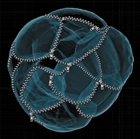
To transform from a loose aggregate of cells into a cohesive spherical tissue, the junctions between the outermost cells of the embryo need to be water-tight, so that the embryo can expand by pumping fluid inside, creating an internal cavity. Until now, it has been unclear what triggers the sealing of early embryos during this transitional phase.
By high-speed imaging, the researchers were able to detect the assembly of the actin rings after the 8-cell stage embryo divides, and track the continued expansion of the rings until they reached the cell-cell junctions. Components that generate tension were then recruited to the actin rings to tightly connect adjoining cells like a zipper.
Detection of these expanding actin rings has been elusive in the past due to technical limitations. The team, co-led by Dr Plachta and Dr Biro, were able to detect the dynamic actin rings and uncover their fundamental importance in embryonic development using a combination of high-resolution imaging with advanced microscopes, fluorescent markers to label actin filaments, and analysis of the biophysical properties of the cells.
“This system is so complex,” Dr Biro said.
“The key was to use an entirely intact model, and to image and probe it non-invasively, something the team members in Singapore are world leaders in.”